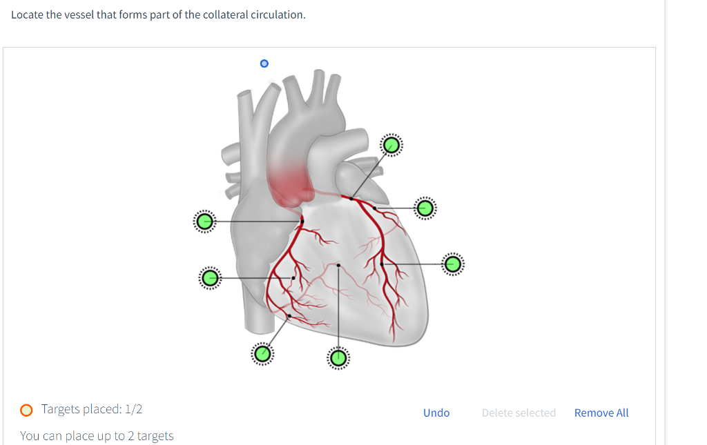
Locate the Vessel That Forms Part of the Collateral Circulation.
Our recent studies showed that VEGF-A places where collateral vessels develop that the protein is indeed expressed on the endothelial surface FGFR-1 is expressed in SMCs of growing collaterals of growing collaterals which explains the mitotic and in fibroblasts of the adventitia 30 and that activity of the endothelium but not of the SMC pro- transgenic myocardial over. The relevance of these collateral arteries is a matter of ongoing debate but increasing evidence indicates a relevant protective role in patients with coronary artery disease.

Circulatory Routes Boundless Anatomy And Physiology
Collateral circulation is generated within a few months around the central vein blocked by a generally winding path usually from an astvene to the choroid.

. The leptomeningeal collateral circulation is a network of small blood vessels in the brain that connects branches of the middle anterior and posterior cerebral arteries with variation in its precise anatomy between individuals. Collateral vessels are extra blood vessels that connect portions of the same artery or link two different arteries. 3Department of Cardiology Saarland University Hospital Saarland Germany Although collateral blood flow after epicardial.
Fullsc O Targets placed. Unanswered Locate the vessel that forms part of the collateral circulation. -part of heart dies.
Up to 10 cash back On magnetic resonance fluid-attenuated inversion recovery FLAIR sequence of patients with AIS we often find serpentine or speckled high signal at several successive levels in the convex sulcus and subarachnoid space of the cerebral hemisphere which was first defined by Cosnard G et al. Pulmonary arteries are the vessels that transfer blood. Obstruction of any one of these pathways will result in blood flow finding new collateral pathways to return to the heart.
Collateral circulation is a network of alternate circulation around a blocked artery or vein via another path such as nearby minor vessels. The collateral circulation can be assessed by different methods. 3 major collateral routes include.
These vessels provide an alternative source of blood supply to the myocardium in cases of occlusive coronary artery disease. Unanswered Locate the vessel that forms part of the collateral circulation. Collateral circulation is present in most tissues and provides protection against ischemic injury caused by ischemic stroke coronary atherosclerosis peripheral artery disease and other conditions and diseases 1.
An anastomosis is the union of the branches of 2 or more arteries supplying the same region of the body. Everyone has collateral vessels but theyre normally small and not used by the circulatory system. As hyperintense vessel sign HVS and.
Fullsc O Targets placed. Collateral circulation-two vessels interconnect to supply the same area-help supply blood to at risk tissue-very small interconnections are normally found among microscopic branches of arteries. The coronary arteries have been regarded as end arteries for decades.
Objectives for Circulation 1. When the coronary arteries narrow to the point that blood flow to the heart muscle is limited coronary artery disease collateral vessels may enlarge and become active. During a stroke leptomeningeal collateral vessels allow limited blood flow when other larger blood vessels provide inadequate blood supply to a.
Collateral circulation is a network of tiny blood vessels and under normal conditions not open. This provides an alternative route for blood flow. 1 The other side of the circle of Willis 2 The posterior cerebral circulation 3 The external carotid artery branches Only 50 of patients have a complete circle of Willis due to variants-these patients will not benefit from the.
Collateral vessels are small blood vessels that connect the aorta the major vessel carrying oxygen-rich blood from the heart to the rest of the body and the main pulmonary artery carrying oxygen-depleted blood from heart to lungs. Understand anastomosis and collateral circulation. 2 the channels of communication between the blood vessels supplying the heart.
02 Undo Delete selected Remove All. These extra vessels are called collateral blood vessels. In the healthy patient blood returns to the heart via classic venous pathways.
COLLATERAL CIRCULATION 1951-11-01 000000 Of the two forms of collateral circulation in which the anterior cerebral artery is involved the basal circulation through the anterior part of the circle of Willis is the more familiar but i n recent years the peripheral type through small anastomosing vessels from one of the major vascular cerebral areas into the other has been. Collateral circulation 1 an alternative route provided for the blood by secondary vessels when a primary vessel becomes blocked. Some areas in your body are supplied by more than one blood vessel.
Almost any vein in the abdomen may serve as a potential collateral channel to the systemic circulation. Up to 10 cash back Schaper W 1993 Collateral circulation heart brain kidney limbs. The aorta is a blood vessel that carries blood from the heart to arteries throughout the body.
Schaper W 1967 Tangential wall stress as a molding force in the development of collateral vessels in the canine heart. Collateral circulation means that more than one artery feeds the capillary bed of an organ. The gold standard involves intracoronary pressure measurements.
PubMed CAS Google Scholar 122. Collateral vessels are abnormal blood vessels that connect the aorta with the pulmonary arteries. These alternate blood circulation routes develop in most people and are usually closed to the flow of blood.
Although significant anatomic variation exists and multiple collateral vessels are often present in the same patient it is a general rule that the collateral pathways. For example there is a. Presence of abnormal collateral vessels appears to be one of the most sensitive 7083 and specific sonographic signs for the diagnosis of portal hypertension.
Vessel That Forms Part of the Collateral Circulation. 02 Undo Delete selected Remove All. However there are functionally relevant anastomotic vessels known as collateral arteries which interconnect epicardial coronary arteries.
Given the central location of so many structures one thing you will find throughout the body are examples of collateral circulation see Figure 123. Arteries that do not form an anastomosis are called end arteries. 1 When blood flow through a vessel or a vascular bed is obstructed due to occlusion as in.

Solved Locate The Vessel That Forms Part Of The Collateral Chegg Com

Circulatory Pathways Anatomy And Physiology Ii

Solved This Question Refers To Ecg Traces In The Figure Chegg Com

Pathogenesis Of The Human Coronary Collateral Circulation Springerlink

Quantification Of Pial Collateral Pressure In Acute Large Vessel Occlusion Stroke Basic Concept With Patient Outcomes Springerlink

Solved Question 18 18 Homework Unanswered Locate The Vessel Chegg Com

Schematics Of Vessel Anatomy And Example Of Angle Of Interaction Aoi Download Scientific Diagram

The Lymphatic System In The Fontan Patient Pathophysiology Imaging And Interventions What The Anesthesiologist Should Know Journal Of Cardiothoracic And Vascular Anesthesia

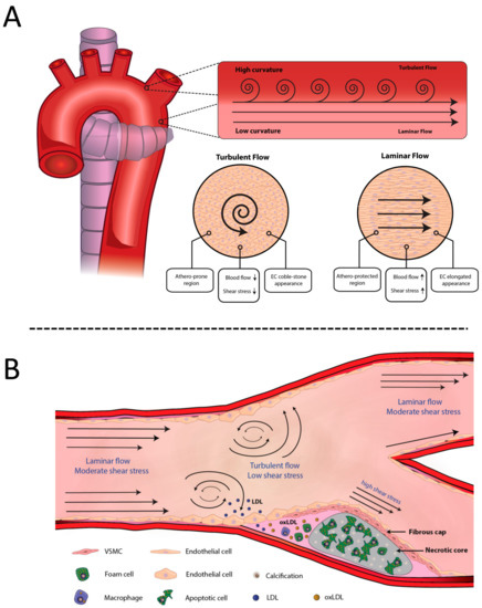
Ijms Free Full Text Pathophysiology Of Atherosclerosis Html

Structure And Function Of Arteries Arterioles Capilaries Venules And Veins Flashcards Quizlet

Coronary Circulation Of The Heart Study Com

Frontiers Pitfalls In The Diagnosis Of Posterior Circulation Stroke In The Emergency Setting Neurology
:watermark(/images/watermark_only_sm.png,0,0,0):watermark(/images/logo_url_sm.png,-10,-10,0):format(jpeg)/images/anatomy_term/arteria-cerebelli-anterior-inferior/thRyBCrBHtmNAGM4Etd2Q_A._inferior_anterior_cerebelli_01.png)
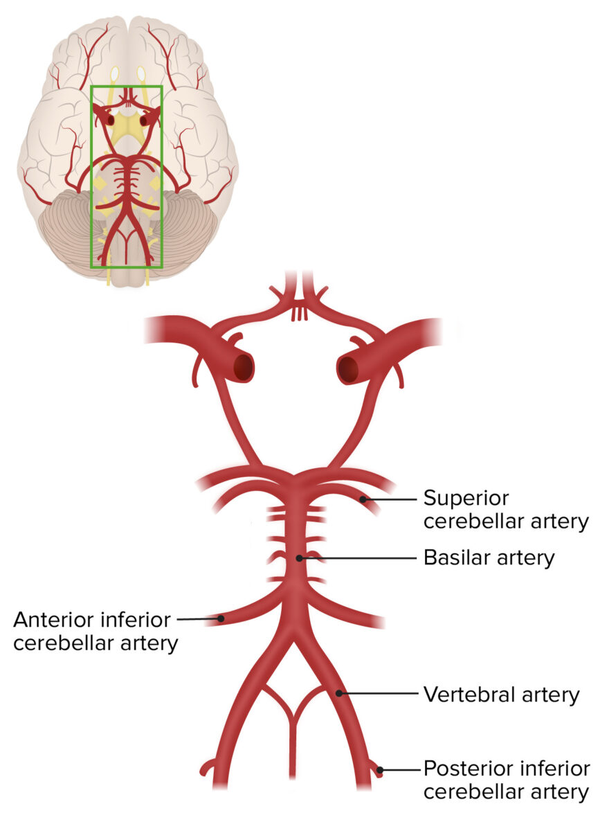
Basilar Artery Anatomy Course And Branches Kenhub

Pedi Cardiology Anatomy Coronary Veins Coronary Arteries Coronary Arteries Coronary Arteries

Organization Of Cerebral Blood Flow Cbf And Collateral Blood Flow Download Scientific Diagram

Endothelial Control Of Cerebral Blood Flow The American Journal Of Pathology

Showing Schematic Diagram Of Anastomosis Of Vessels Around Ankle And Download Scientific Diagram


Comments
Post a Comment